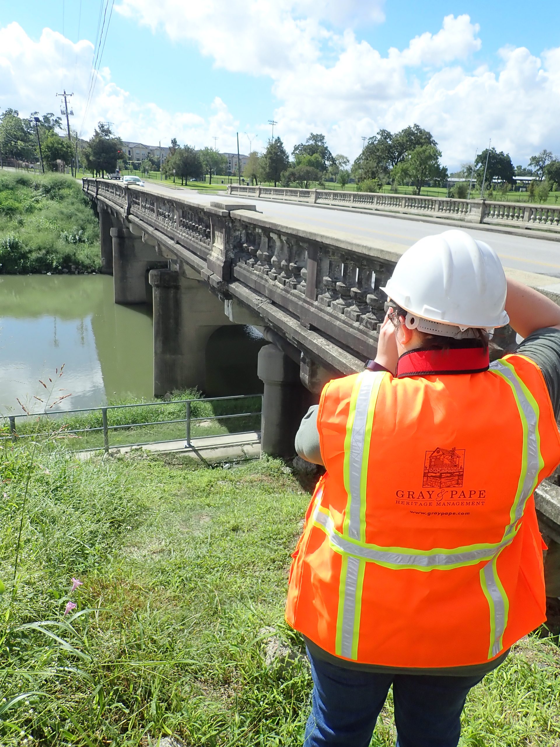
Gray & Pape serves our clients, our profession, and our nation's cultural resources by providing the highest quality cultural resources management services.
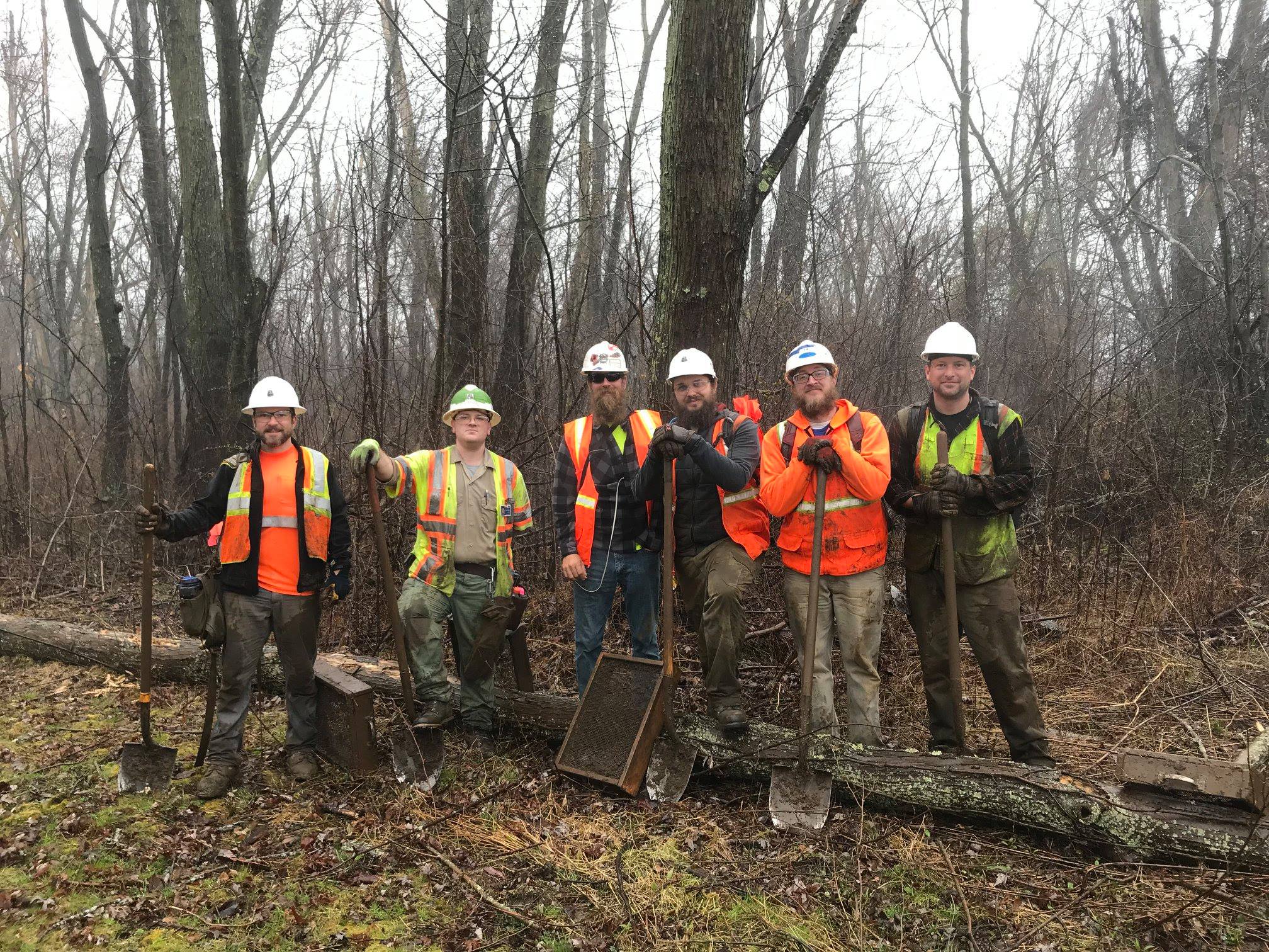
Archaeology
Cultural Heritage
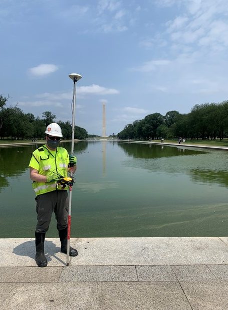
History Architecture
Maritime Heritage
Who We Are
Gray & Pape, Inc., is a small-business enterprise, with over 35 years’ experience specializing in a national and international practice that delivers cultural heritage management services to public and private industry clients.


